
A 19-year-old construction worker was brought into hospital having been found unconscious at the bottom of a stairwell at work. He had been unconscious for 45 mins according to CCTV. He was usually well, but he’d had episodes of vomiting, and according to friends and family had been a little vague for 2 weeks leading up to this episode.
By the time he got to hospital he was awake and talking but confused and unsure of what had happened.
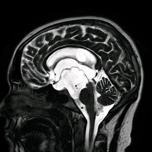
A CT and subsequently an MRI of the brain (Fig 1) showed very large ventricles. Ventricles are fluid filled spaces within the brain we are born with.
Cerebrospinal fluid (CSF) is produced in the ventricles and circulates around the brain and spinal cord. In this patient’s case a large cyst was found within his ventricular system obstructing normal CSF communication and causing a back-log of CSF within the ventricles, which again causes the ventricles to enlarge.
The patient had surgery through an endoscope – a tube approx. 4mm in diameter with a camera at the end. This allows us to directly observe the structures. The endoscope has a port where small micro instruments can be passed in through the endoscope to perform surgery. At the time of surgery, we saw the cyst wall which was unusually thick (Fig 2). We used small grabbing tools, scissors and coagulation to make an opening into the cyst (Fig 2). Opening the cyst up causes the cyst to collapse and normal CSF flow in restored.
He is very happy with the result of his surgery and he was discharged from hospital 3 days after surgery. He has resumed normal life. 3 years later he has not had any further symptoms and repeat MRI’s show the cyst remains collapsed and the ventricles are returning to a normal size (Fig 3).